Overview of Building a digital ant gallery, from the ground up
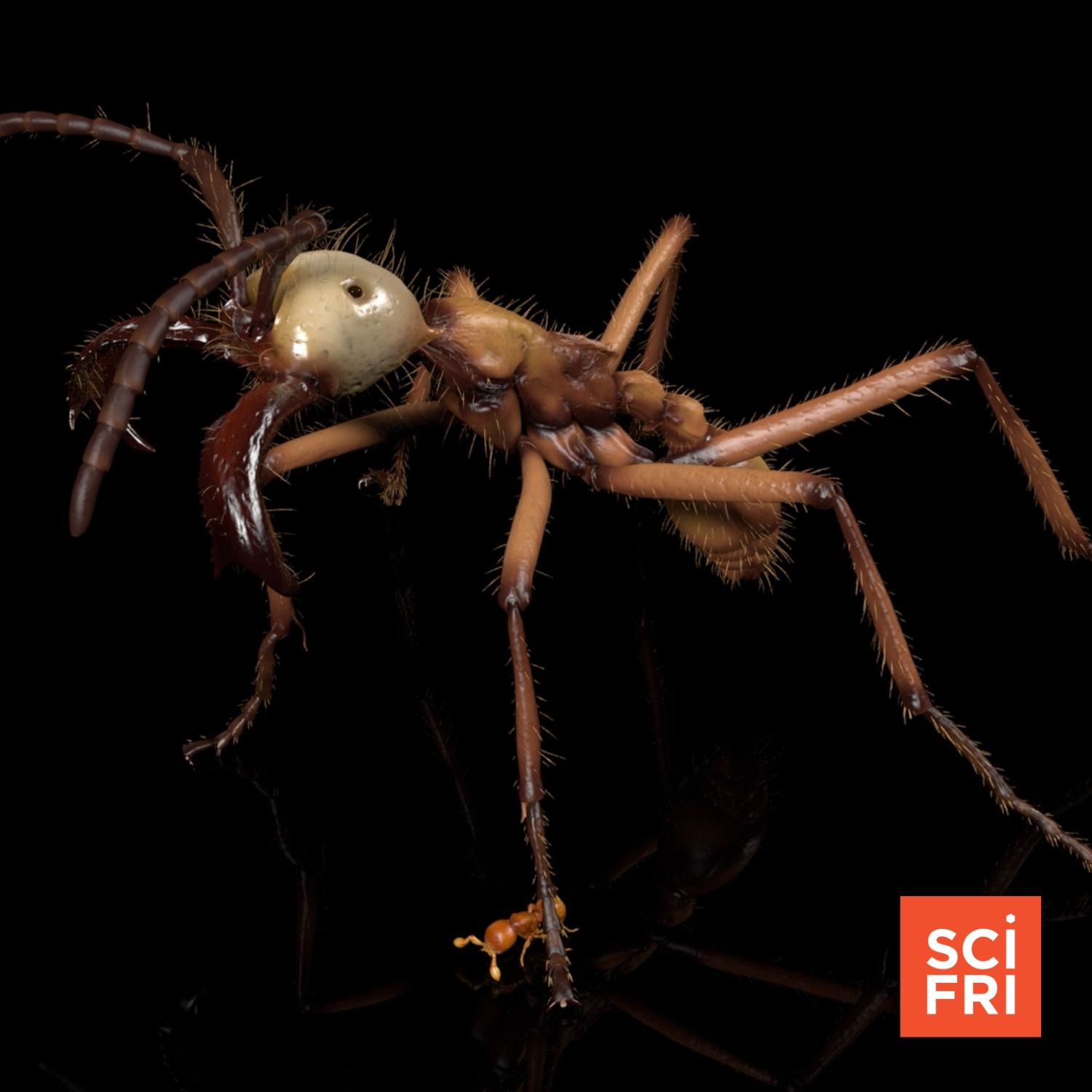
This Science Friday episode (host Flora Lichtman) interviews Dr. Julian Katsuka about AntScan — a large, open digital library of high‑resolution 3D x‑ray (micro‑CT) scans of ants. AntScan contains scans of over 2,000 specimens from more than 700 species, revealing external and internal anatomy at micrometer resolution. The project aims to accelerate research on ant form, function, and evolution while also enabling public engagement, animation/game use, and development of AI tools for automatic annotation.
Key points and takeaways
- AntScan produced high‑resolution 3D reconstructions of >2,000 ants across >700 species using synchrotron micro‑CT.
- The project resolves external fine details (hairs, mandibles) and internal anatomy (muscles, guts, nervous tissue) — voxel size down to 1.22 µm.
- Imaging speed: roughly 30 seconds per ant for scanning plus ~30 seconds for data transfer; throughput increased by using a robot to exchange samples (50 at a time during the project).
- Hospital CT scanners lack the needed resolution — AntScan used a synchrotron light source and high‑speed cameras to achieve micro‑CT quality.
- Data are made freely accessible for scientists, educators, artists, game developers, and the public.
- Main research applications: comparative morphology, evolution of mouthparts and other traits, biomechanics (e.g., ant strength).
- Follow‑on goals: annotate datasets, train AI models to segment tissues and automate trait extraction, and scale the approach to other invertebrates.
How AntScan works (method overview)
- Imaging modality: synchrotron X‑ray micro‑CT (high energy, high resolution).
- Workflow highlights:
- Samples mounted and swapped by a robotic system to increase throughput.
- High‑speed cameras capture many projection images; mathematical reconstruction yields 3D grayscale image volumes (tomography).
- Volumes can be segmented, colored, and rendered for analysis or visualization.
- Performance:
- Scan time per specimen: ~30 seconds; transfer ~30 seconds.
- Resolution: voxel = 1.22 micrometers (enough to see hairs and individual muscle fibers).
- Compared to standard lab micro‑CT: previous workflows could take 8–12 hours per insect.
Scientific and practical uses
- Research:
- Large‑scale comparative studies of morphology and trait evolution (e.g., mandible diversity and function).
- Internal anatomy studies (musculature, nervous tissue) enabling biomechanics and functional analyses.
- Integrating scans with phylogenies to test evolutionary hypotheses.
- Technology & AI:
- Training segmentation models to distinguish exoskeleton vs. muscles vs. nervous tissue at scale.
- Automating measurement and trait extraction across thousands of specimens.
- Public engagement & media:
- High‑quality renders for museum exhibits, educational resources, and science communication.
- Assets for 3D artists, animators, and game developers to use accurate, detailed insect models.
- Outreach: making tiny biodiversity visible at human scales helps people appreciate morphological diversity.
Notable quotes and highlights
- “The biomass of all ants equals or surpasses that of like all humans.” — emphasizes ecological abundance and importance.
- “You can resolve the delicate hairs… and individual muscle fibers.” — on the level of anatomical detail captured.
- “They look like aliens… it’s beautiful and it’s an acquired taste.” — about the aesthetic and uncanny quality of the scans.
- Practical bottleneck solved: reducing per‑specimen scan time (and using a robot) enabled scaling from single scans to thousands.
Action items / recommendations
- View the gallery and sample images at Science Friday: sciencefriday.com/ants (episode provides curated visuals).
- For researchers: consider downloading the open dataset for morphological, phylogenetic, or biomechanical analyses and plan annotation/segmentation pipelines for AI training.
- For educators/creators: use the 3D scans as resources for exhibits, teaching materials, animations, or games that want accurate insect models.
- For funders/collections: support scaling this pipeline to other invertebrate groups to expand a digital library of small animal anatomy.
Credits and people
- Guest: Dr. Julian Katsuka — postdoc at the Smithsonian Museum of Natural History; did this work as a PhD student at Okinawa Institute of Science and Technology.
- Host: Flora Lichtman, Science Friday (WNYC Studios).
- Episode produced by Charles Berquist.
If you want a quick visual hit, the episode points to the online gallery — the scans are striking and worth inspecting to appreciate ant diversity up close.